Researchers at the Institute for Basic Science (IBS) managed to observe the movement of molecules stored inside a graphene pocket without the need to stain them. This study opens the door to using graphene for observing the dynamics of life building blocks like proteins, DNA and more.

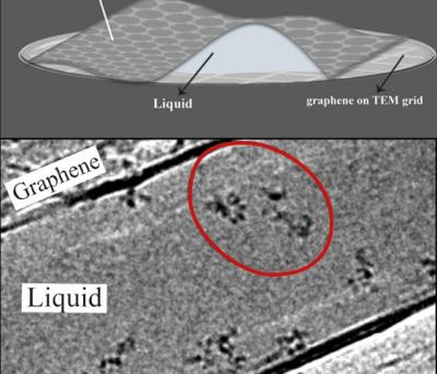
Due to its operating mechanism, electron microscopy is known to be suitable for visualizing inert, dead samples, while living material needs to be chemically locked in place. IBS scientists broke this rule and visualized non-fixed chains of atoms, called polymers, swimming in a liquid inside graphene pockets. These consist of 3-5 graphene layers on the bottom and two on top. The sheets are impermeable to small molecules, and also prevent the electron beam from instantly harming the sample: the scientists had an average of 100 seconds to admire the dynamic movement of individual polymer molecules, before these were destroyed by the electron beam. During these valuable seconds, molecules change position, rearrange or "jump around".
The team explained that previously, it was necessary to stain samples with metal or dye molecules to make them visible inside the graphene pocket. Metal has high reflexibility, so it can be used to get good images. However, the chemical bonds between the sample and the metal or dye changes the characteristics of the sample molecule. In this study, the graphene pocket is thin enough that its content can be observed in real time without any staining.
In particular, the scientists worked with two polymers: one with sulfur, polystyrene sulfonate, and one without, polyethylene oxide. This allowed them to show that the contrast under the microscope comes from the polymer structure - made of carbon and hydrogen atoms - rather than from the sulfur. "Most molecules produced by living organisms have a backbone made of carbon and hydrogen, and that's why we hope to extend this research to the study of interactions between DNA and proteins," explains the first co-author. Moreover, since the scientists used a standard electron microscope, they expect that this technique will be used in other laboratories.