Researchers at the University of Zurich in Switzerland have managed to use graphene to deliver the world's first picture of a single protein. Taking pictures of proteins help scientists understand proteins' structure and functions, which is vital for treating diseases in which proteins go wrong, like Alzheimer’s. But imaging methods such as X-ray crystallography or cryo-electron microscopy rely on averaging readings from millions of molecules (granting a blurry view), since illuminating molecules with X-rays or high-energy electrons can damage the protein.
The scientists started by spraying a solution of the proteins onto a sheet of graphene, fixing the proteins in place. Then they placed this under an electron holographic microscope, which uses interference patterns between electrons to produce an image. While this kind of instrument relies on low-energy electrons that don’t damage the protein, it is also less able to penetrate through to the microscope’s detector - which is where graphene comes in handy as the substrate can be made thin enough to have the electrons passing through.
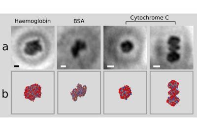
The team tested their method on a range of protein molecules, all just a few nanometres in size, such as the haemoglobin found in red blood cells. The results sat well with molecular models derived from X-ray crystallography (as can be seen in the image), suggesting the images are accurate. Now they plan to take pictures of other molecules that can’t be imaged with existing techniques, and hope eventually to contribute to new medical treatments.