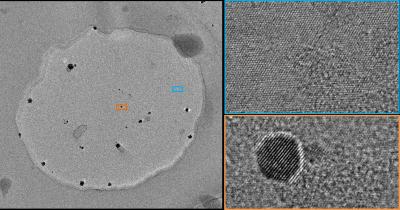
Researchers at The University of Manchester and the NGI have shown how graphene and boron nitride can be used for observing nanomaterials in liquids, by creating a ‘petri-dish’ of sorts.

Scanning / transmission electron microscopy (S/TEM) is one of only few techniques that allows imaging and analysis of individual atoms. However, the S/TEM instrument requires a high vacuum to protect the electron source and to prevent electron scattering from molecular interactions. Several studies have previously revealed that the structure of functional materials at room temperature in a vacuum can significantly different from that in their normal liquid environment. So, it is important to be able to study the structure at the required state.
These engineered graphene liquid cells (EGLC) are built from 2D material-building blocks: they consist of a boron nitride (BN) spacer drilled with holes (where the liquid is contained) and encapsulated with graphene on both sides.
The team explains that graphene is the ultimate window material - strong enough to protect the sample from a high vacuum environment, but at the same time thin enough that the resolution of the electron beam is not compromised. Lead author Daniel Kelly said: Unlike some previous designs our graphene liquid cells allow us to image the atoms for many minutes. We were even able to resolve individual atoms in water and observe them dancing under the electron beam.
The researchers also demonstrated that these new graphene liquid cells enable an order of magnitude improvement in the quality of elemental analysis in liquid cells. They studied the deposition of a 1nm shell of iron on gold to grow core-shell nanoparticles. This new ability to monitor tiny concentrations over such small length-scales is a necessity for the increasingly complex chemical structures of high-performing nanocatalysts.