Researchers create graphene-integrated bioelectronic mesh for tracking multimodal excitation-contraction dynamics in cardiac microtissues
Researchers at the University of Massachusetts and Massachusetts Institute of Technology (MIT) have successfully built a tissue-like bioelectronic mesh system integrated with an array of graphene sensors that can simultaneously measure both the electrical signal and the physical movement of cells in lab-grown human cardiac tissue.
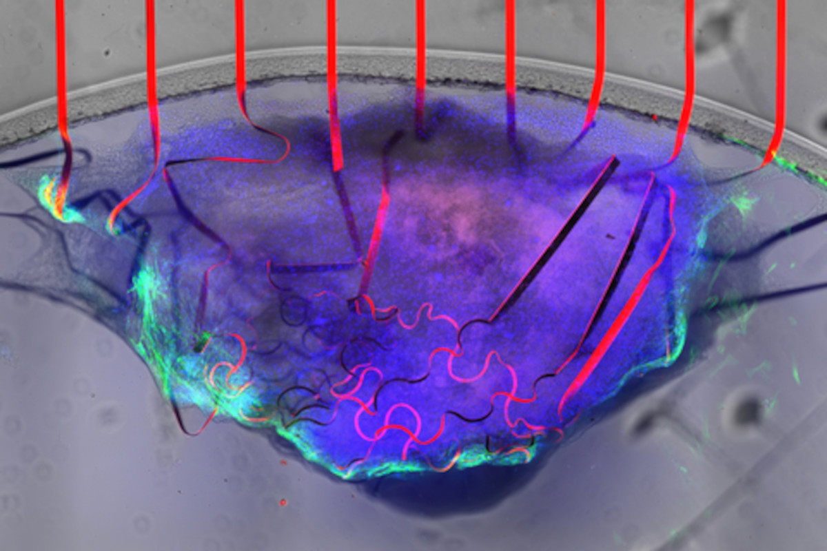
A bioelectronic mesh, studded with graphene sensors (red), can measure the electrical signal and movement of cardiac tissue (purple and green) at the same time. Image credit: UMass Amherst
The tissue-like mesh can grow along with the cardiac cells, allowing researchers to observe how the heart’s mechanical and electrical functions change during the developmental process. The new device can be extremely useful for those studying cardiac disease as well as those studying the potentially toxic side-effects of many common drug therapies.